
DNA methylation biomarkers
AI-driven lung cancer detection using epigenetic signatures.
This conference study evaluates whether methylation patterns across EGFR, PD-L1, SHOX2, RASSF1A, and PTGER4 can support non-invasive lung cancer detection across lung adenocarcinoma and lung squamous cell carcinoma cohorts.
The core lesson is that individual biomarkers were not enough on their own. The stronger signal emerged after feature engineering and a gradient boosting model combined weak molecular patterns into a more stable diagnostic classifier.
EGFR
PD-L1
SHOX2
RASSF1A
PTGER4
gradient boosting
TCGA LUAD
TCGA LUSC
EGFR methylation
PD-L1 methylation
SHOX2 methylation
RASSF1A methylation
PTGER4 methylation
Gradient boosting
Python
ROC and heatmaps
TCGA LUAD
TCGA LUSC
EGFR methylation
PD-L1 methylation
SHOX2 methylation
RASSF1A methylation
PTGER4 methylation
Gradient boosting
Python
ROC and heatmaps
What it does
Converts methylation biomarker measurements into a validated classifier.
Assemble lung cancer methylation cohorts
Analyzes LUAD and LUSC methylation data across thousands of tumor and normal samples.
Engineer a compact biomarker feature set
Reduces 63 initial variables to 9 engineered features after quality control and preprocessing.
Train a gradient boosting classifier
Uses hyperparameter tuning and stratified validation to combine weak individual methylation signals.
Evaluate clinical-facing behavior
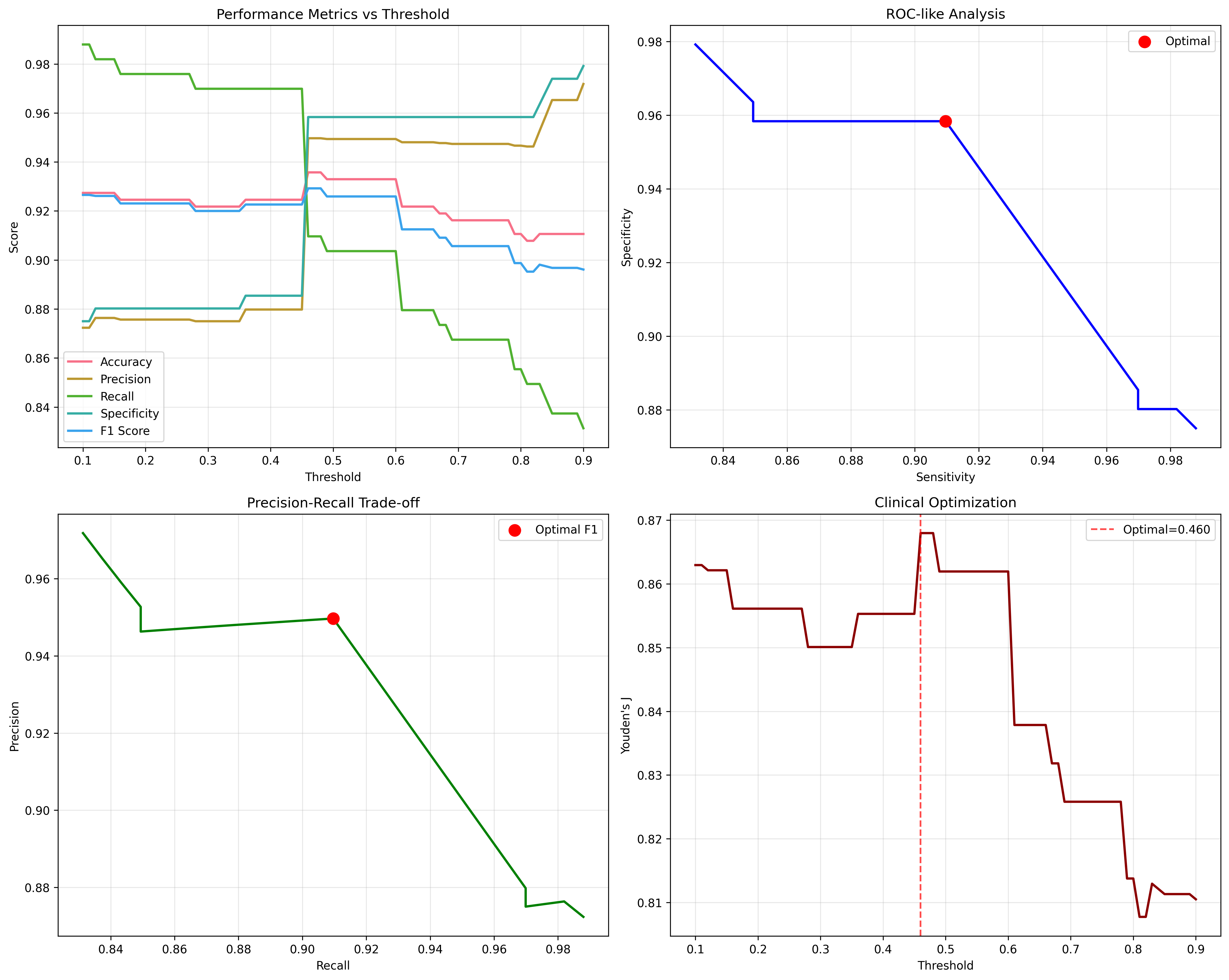
Reports ROC-AUC, accuracy, precision, recall, cross-validation stability, and threshold behavior.
Why it matters
The model is strongest because it learned from biomarker limitations.
RASSF1A showed the strongest individual signal among the five markers, but the single-marker results were not enough for a strong diagnostic tool. The project became more compelling when it showed why ensemble learning was necessary: the model combined subtle, imperfect methylation patterns into a stronger classifier.
Dataset scale
Screened 7,240 methylation samples, with 2,385 final processed samples after feature engineering.
Independent test performance
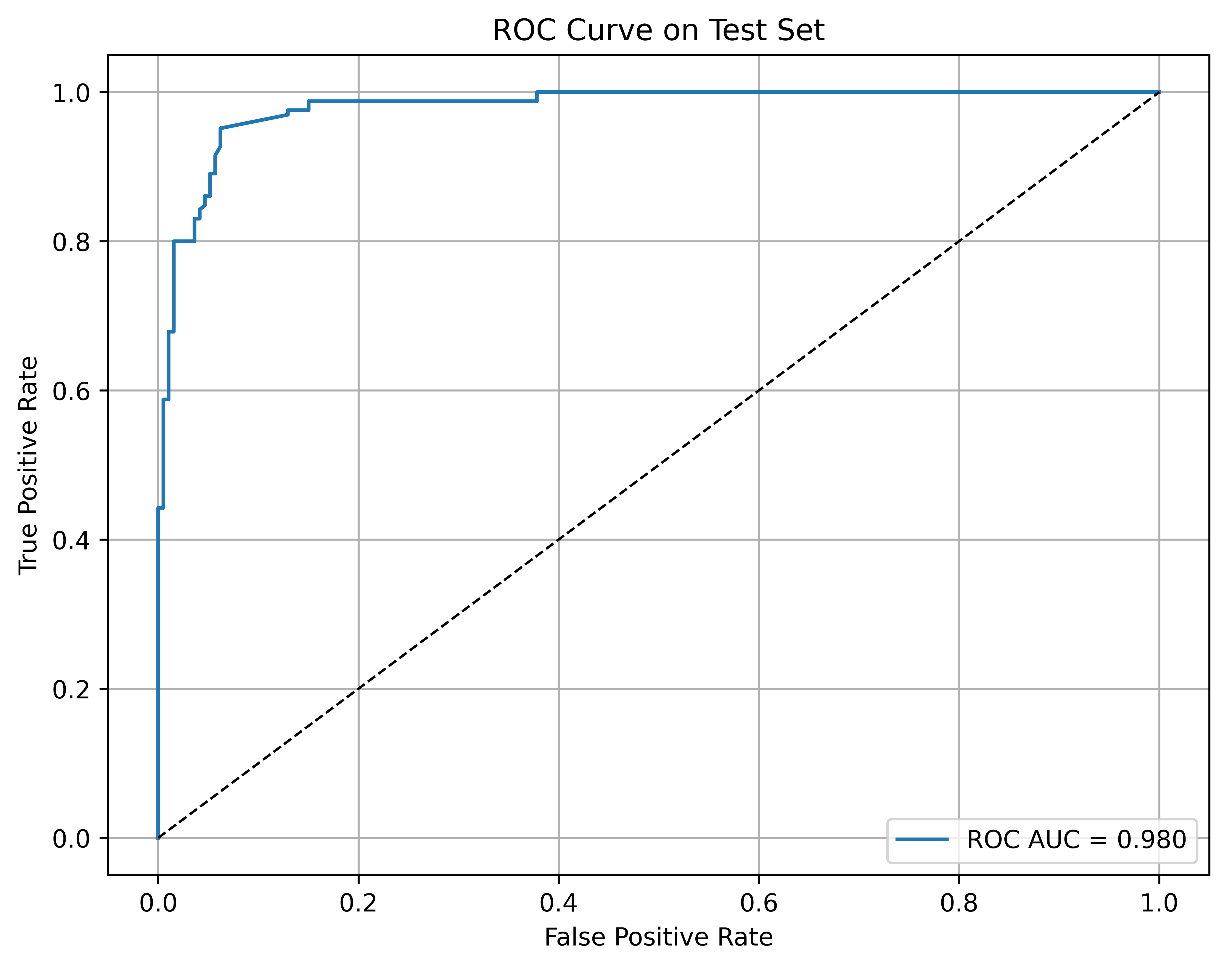
Reported 0.983 ROC-AUC, 92.7% accuracy, 87.6% precision, and 98.2% recall on the test set.
Cross-validation stability
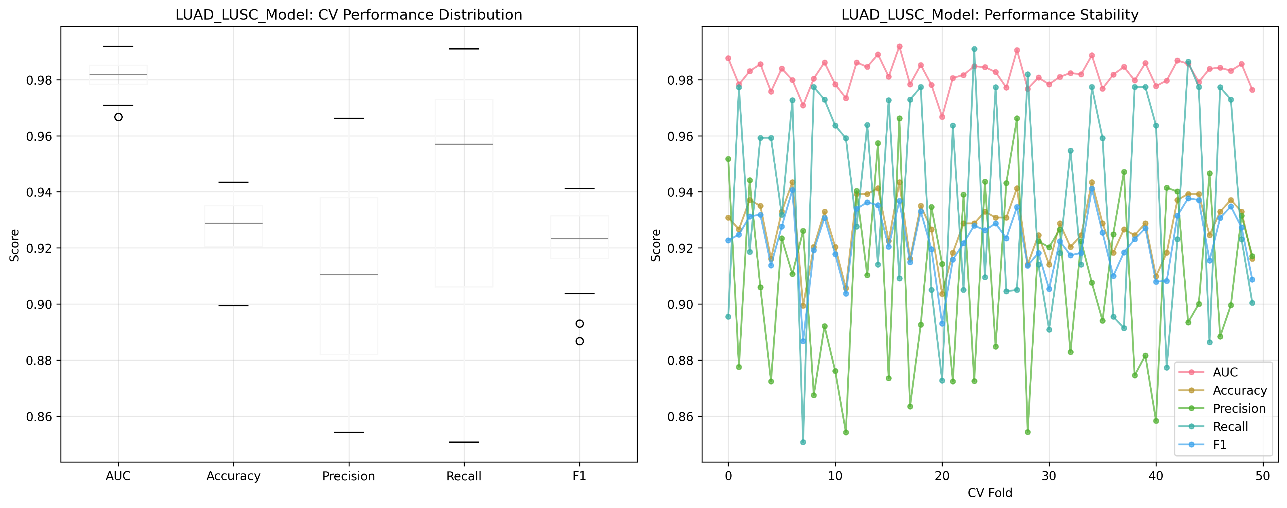
Reported cross-validation ROC-AUC of 0.982 +/- 0.005, suggesting the model was not dependent on one split.
Biological interpretation
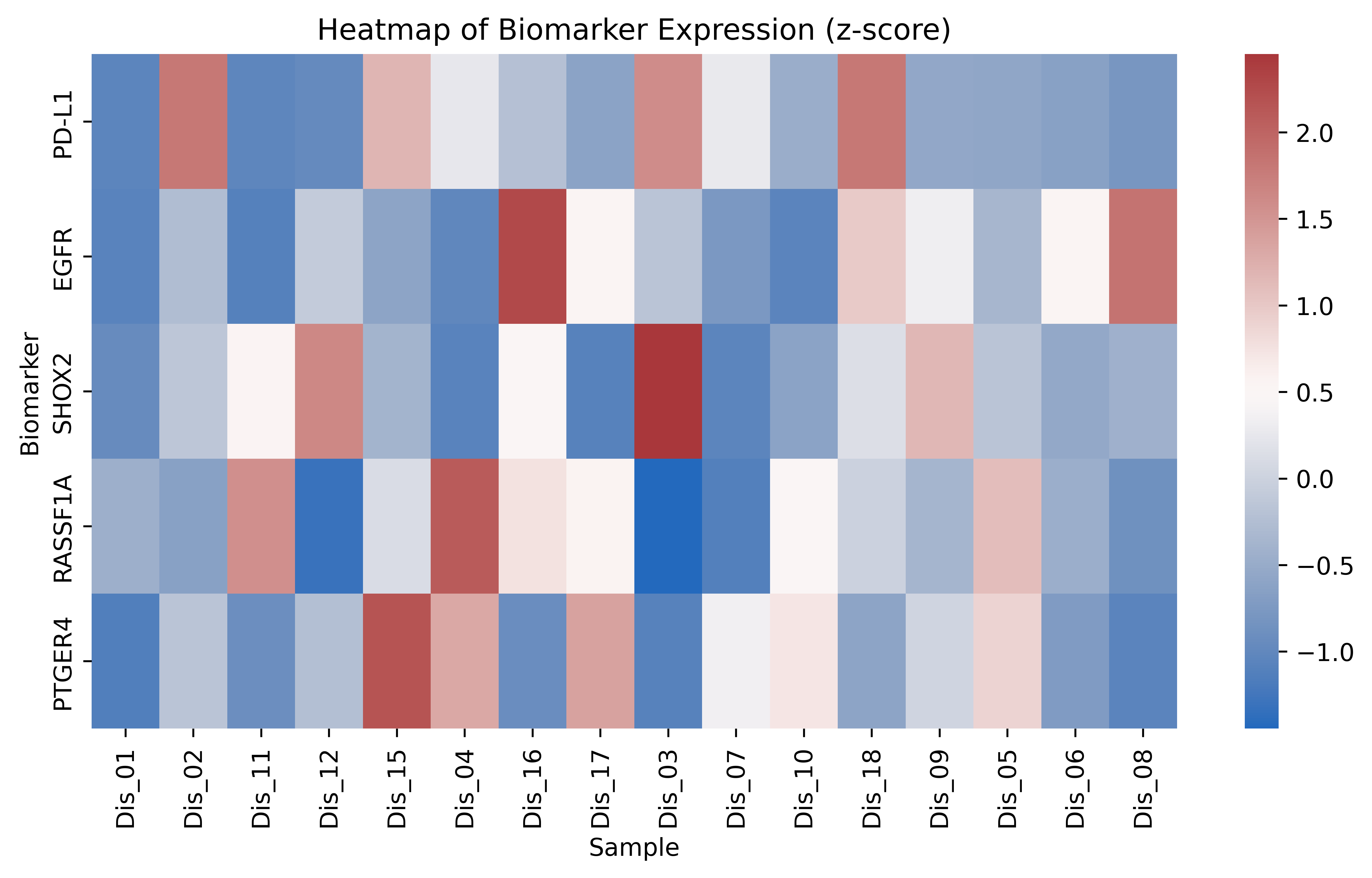
Stage-dependent methylation patterns, especially for RASSF1A and SHOX2, support biological plausibility.
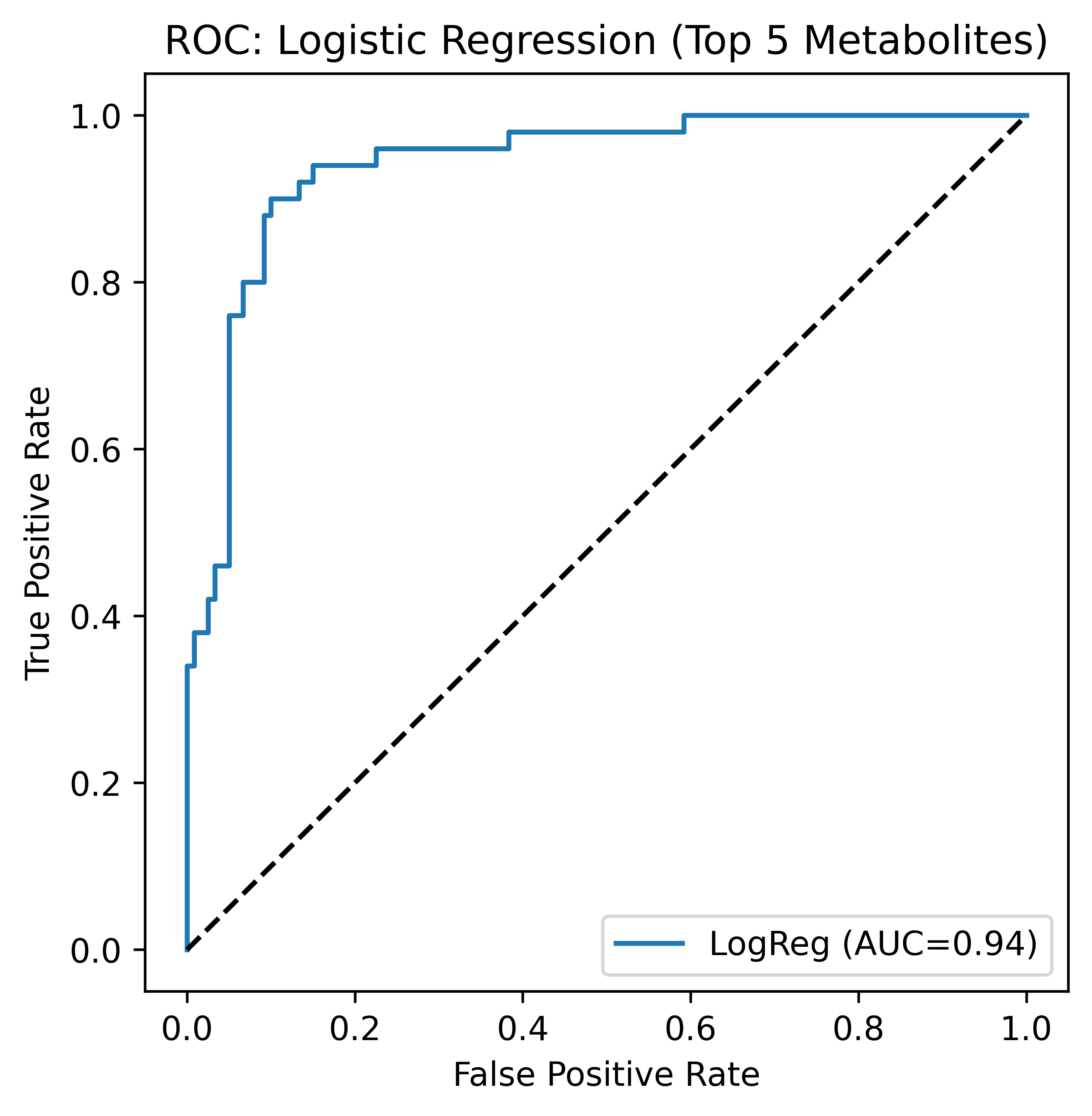
Figures
Selected evidence from the methylation biomarker study.
Data sources and tools
Where the methylation data came from, and how it was modeled.
The methylation project uses public lung cancer molecular cohorts and keeps the method focused on reproducible preprocessing, compact biomarker features, gradient boosting, and interpretable validation plots.
 TCGA LUAD / LUSC
Public lung adenocarcinoma and lung squamous cell carcinoma molecular datasets used for tumor-normal methylation analysis.
 Public cancer data portals
Open molecular-data portals used for dataset discovery, cohort cross-checking, and clinical label context.
 Python
Preprocessing, model training, evaluation, and reproducible analysis.
 pandas
Methylation tables, labels, feature matrices, and cohort organization.
 NumPy
NumPy
Numerical transformations and matrix operations for biomarker features.
 scikit-learn
Gradient boosting, validation metrics, and threshold evaluation.
 Matplotlib / Seaborn
ROC curves, biomarker heatmaps, validation plots, and threshold figures.
Manuscript status
Conference manuscript details available by request.
The public page summarizes the method, results, and selected figures without exposing private submission files. The manuscript can be shared directly by email when appropriate.