
MiNAE
Mutation-independent Nucleosome Architecture Evaluation
MiNAE is a cfDNA fragmentomics framework for hepatocellular carcinoma detection. Instead of searching for tumor mutations, it studies the physical size patterns of cell-free DNA fragments and asks whether those patterns distinguish liver cancer from chronic liver disease controls such as cirrhosis and hepatitis B.
The analysis uses public FinaleDB fragment-coordinate files and keeps the claim deliberately bounded: MiNAE is not a clinical diagnostic. It is a patient-level validation and auditing framework designed to show what cfDNA fragment-size architecture can and cannot prove from public coordinate-level data.
176 patients
HCC vs cirrhosis/HBV
ROC/PR curves
ablation study
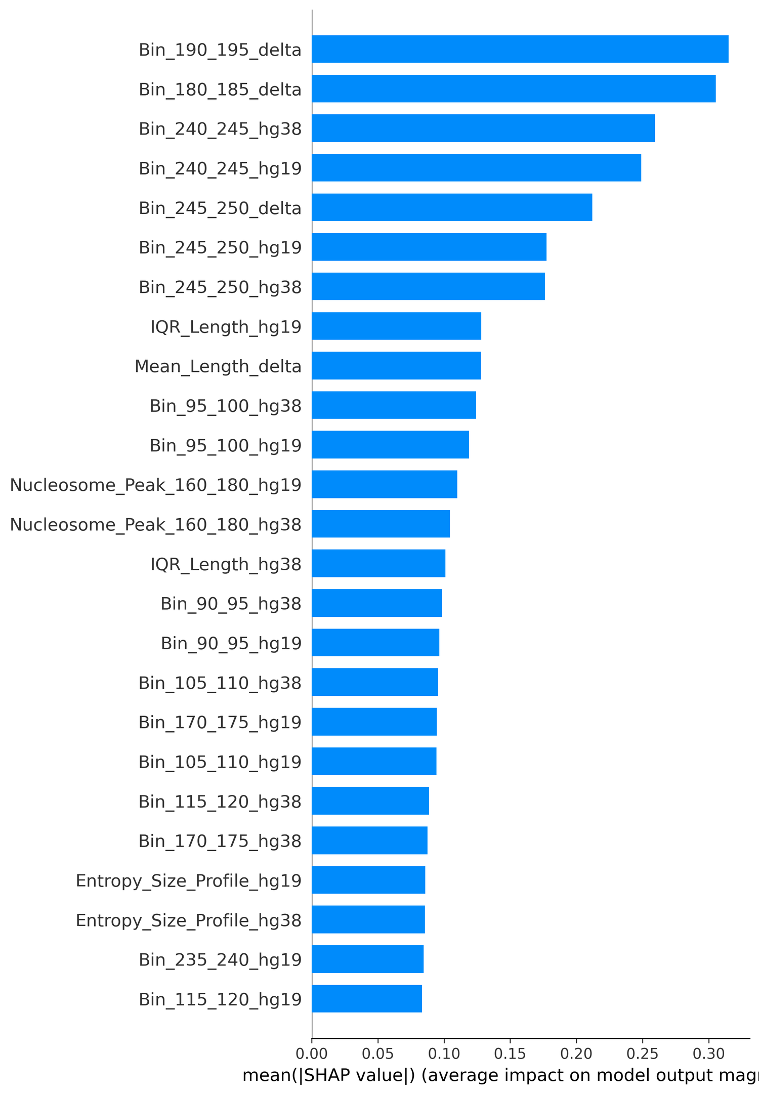
SHAP
technical audit
FinaleDB liver cfDNA
HCC cases
Cirrhosis controls
Hepatitis B controls
hg19 / hg38 split audit
fragment lengths 50-250 bp
ROC and PR curves
Foda / Annapragada reference
Python
Linear SVM
FinaleDB liver cfDNA
HCC cases
Cirrhosis controls
Hepatitis B controls
hg19 / hg38 split audit
fragment lengths 50-250 bp
ROC and PR curves
Foda / Annapragada reference
Python
Linear SVM
What it does
From compressed fragment coordinates to leakage-aware patient predictions.
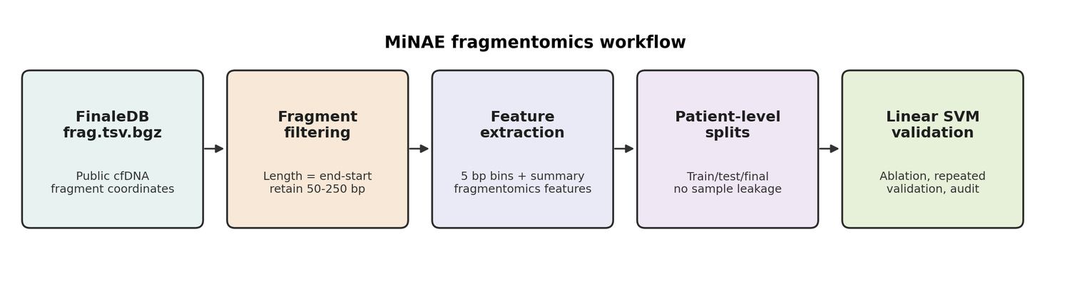
Ingest fragment-coordinate data
Reads public FinaleDB cfDNA fragment-coordinate files from liver cancer, cirrhosis, and hepatitis B sample groups.
Calculate fragment lengths
Computes the physical DNA fragment length as genomic end minus genomic start, then filters likely artifacts.
Build size-profile features
Turns millions of raw rows into patient-level normalized fragment-size fingerprints.
Validate with clinical controls
Tests whether the model separates HCC from chronic liver disease rather than from perfectly healthy samples.
Prevent reference-build leakage
Groups hg19 and hg38 versions of the same biological sample before splitting so one patient cannot appear in both training and validation.
Audit the signal
Compares feature strategies, repeated splits, permutation tests, external fragment-length replication, and technical negative controls.
Validation logic
The project became stronger when it stopped chasing one lucky score.
The most important improvement was moving beyond a single train-test split. MiNAE now includes ablation studies, repeated validation, PR-AUC, confusion matrices, SHAP interpretation, external replication audits, and explicit technical-confounder checks.
Clinical comparator
The final cohort used 89 HCC cases and 87 chronic liver disease controls, including 66 cirrhosis and 21 hepatitis B samples.
Ablation study
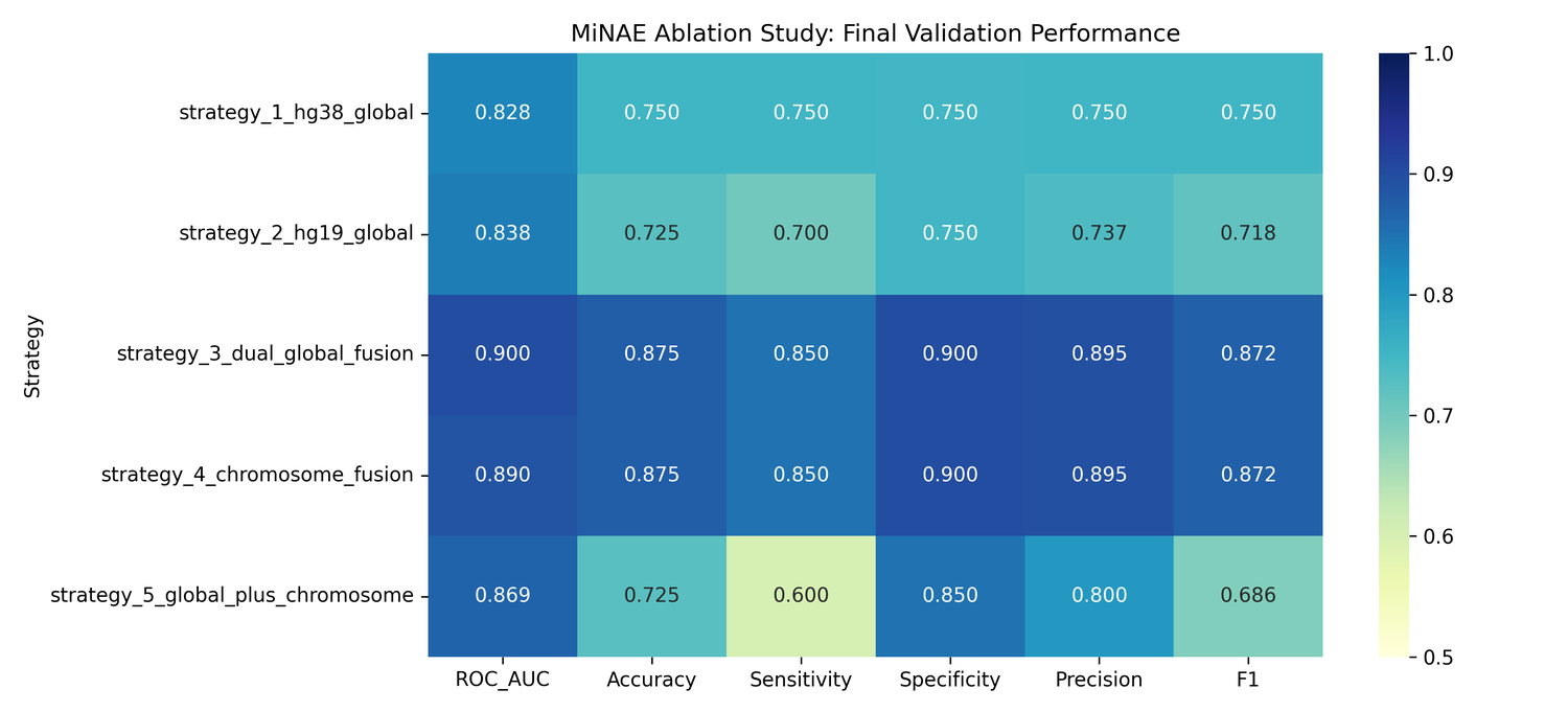
Compares hg38 global, hg19 global, dual-reference fusion, chromosome-focused features, and combined strategies.
Interpretability
SHAP and selected-feature heatmaps help show which fragment-size features drive the classifier.
Honest limitation
External datasets were useful for fragmentomics replication and auditing, but not a perfect HCC reproduction cohort.
Research value
The useful part is not only the score. It is the validation discipline.
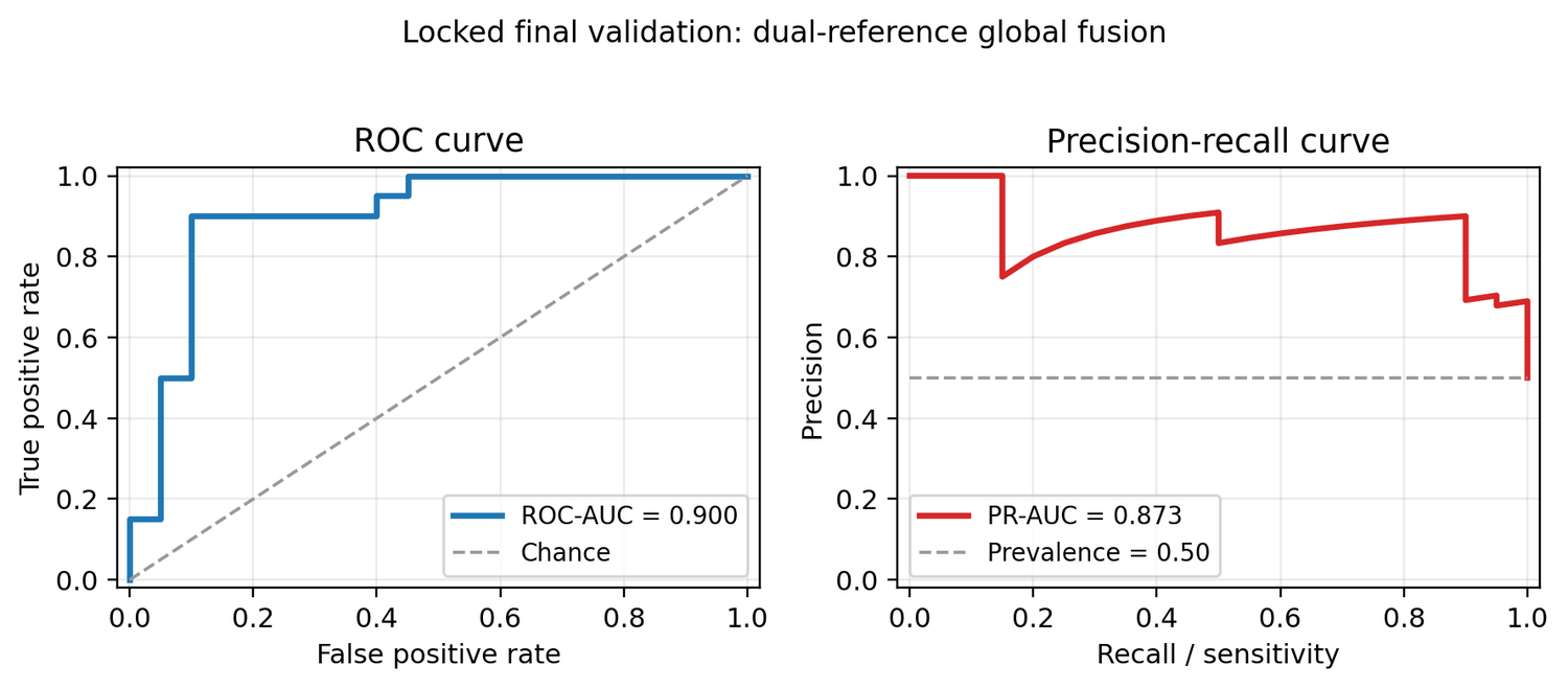
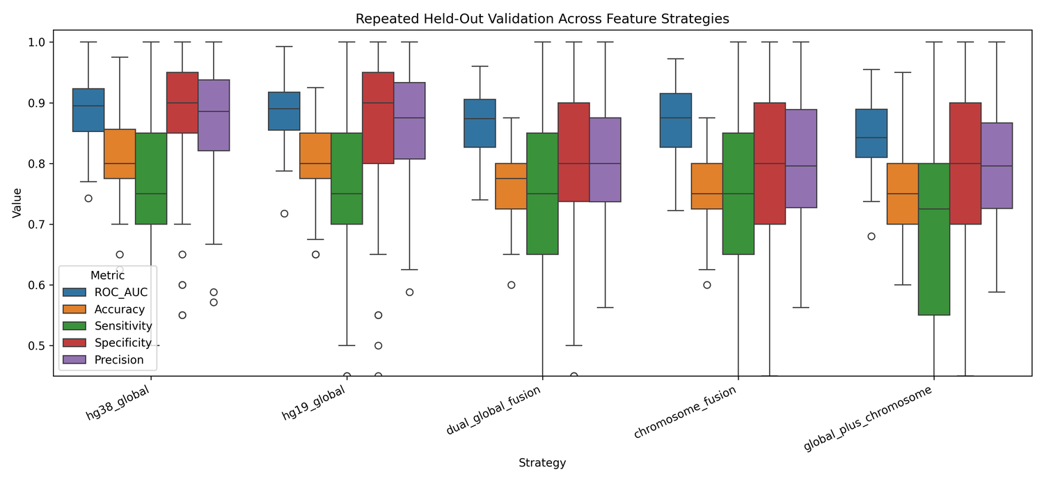
MiNAE is strongest when read as an example of how a public biomedical dataset should be tested. The locked split showed strong HCC separation, but the repeated validation told a more careful story: simpler hg38 global fragmentomics was the most stable strategy across randomized patient-level splits.
What worked
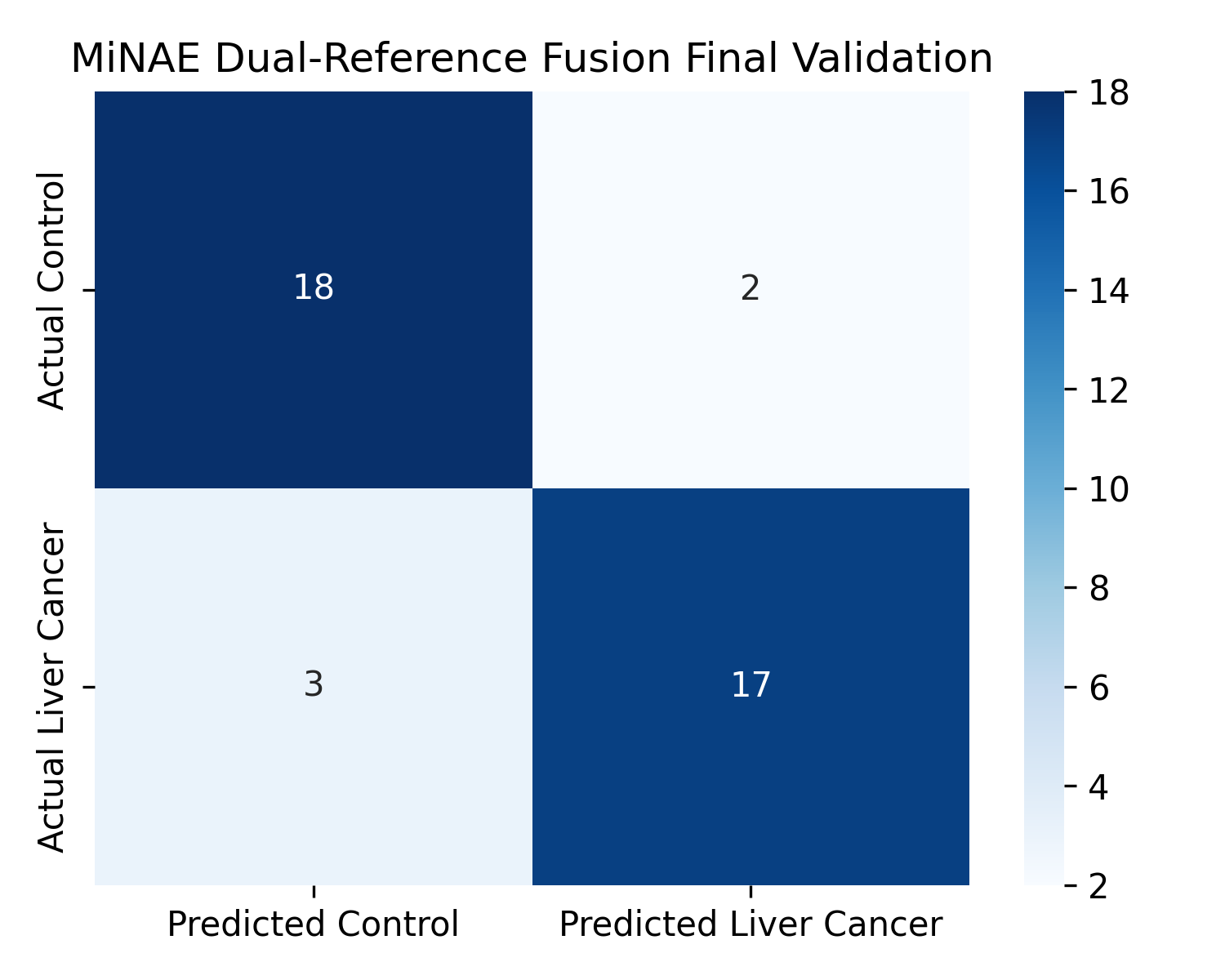
Dual-reference global fusion reached 0.900 ROC-AUC, 0.873 PR-AUC, 0.875 accuracy, 0.850 sensitivity, and 0.900 specificity on the locked final validation set.
What generalized better
Across 100 repeated validations, the simpler hg38 global model was more stable, with mean ROC-AUC 0.889 and mean PR-AUC 0.912.
What reviewers would ask
A technical negative-control model was also predictive, so the website and manuscript frame MiNAE as a retrospective audit, not a deployable clinical test.
What it teaches
Fragmentomics appears measurable in public data, but credible biomedical ML needs patient-level splitting, ablation, PR curves, external checks, and confounder analysis.
Interactive evidence
A 3D surface for the ablation and validation landscape.
This interactive surface is a website visualization inspired by classic 3D performance landscapes. It maps MiNAE feature strategies against validation metrics so the result is seen as a shape across evidence layers, not as a single isolated AUC number.
MiNAE evidence surface
What the surface is showing
The high ridge appears where dual-reference and chromosome-aware strategies performed strongly on the locked split. The flatter region shows why repeated validation mattered: more features did not always make the model more stable.
- Drag the surface to rotate it.
- Hover the surface to read the nearest strategy, metric, and score.
- Use the 2D ROC, PR, ablation, and confusion figures for manuscript work.
Hover the surface
Figures
Selected visual evidence from the MiNAE analysis.
Data sources and tools
Where the MiNAE data came from, and what processed it.
FinaleDB was the primary downloadable source for coordinate-level cfDNA fragments. External HCC fragmentome studies were used as reference and audit context, while the model itself stayed transparent: scientific Python, patient-level splitting, interpretable metrics, and reviewer-facing figures.
 FinaleDB
Primary source for public de-identified cfDNA fragment-coordinate files, including liver cancer and chronic liver disease groups.
 Foda / Annapragada HCC fragmentomes
External liver-cancer fragmentome reference used to ground the audit and related-work framing; raw controlled-access data should not be overstated as open.
 Python
Feature extraction, validation scripts, and analysis workflow.
 pandas
Patient-level feature tables and matrix handling.
 NumPy
NumPy
Numerical feature normalization and array operations.
 scikit-learn
Linear SVM, feature selection, metrics, and repeated validation.
 SHAP
SHAP
Feature-level interpretability for fragment-size signals.
 Matplotlib / Seaborn
ROC, PR, heatmap, confusion-matrix, and validation figures.
 Google Colab
Notebook-style experimentation and portable model runs.
Public manuscript status
Draft available by request, not publicly posted yet.
Because MiNAE is being prepared for conference submission, I am not publicly linking the full draft manuscript here. A project page with methods, figures, and an email request link is safer until acceptance or formal preprint posting.